When you first look at a dental X-ray, it all comes down to a simple principle: dense materials appear white, while softer tissues and problem areas like decay show up as darker shadows. It’s this fundamental contrast that helps us see what’s really going on below the surface.
Your First Look at Understanding Dental X-Rays

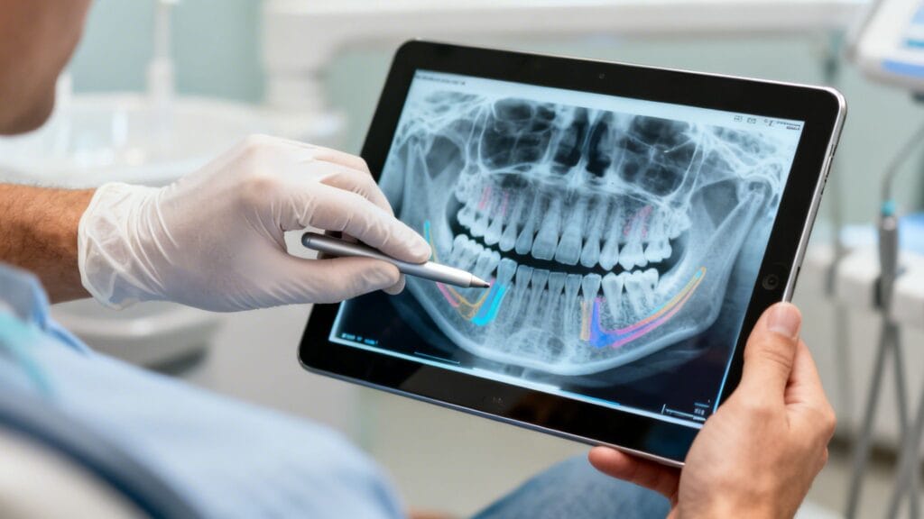
Does looking at your dental X-rays feel a bit like trying to solve a puzzle? Those black, white, and gray images can seem a little intimidating, but they are one of our most important tools for spotting problems that are completely invisible to the naked eye.
The concept is actually pretty straightforward. As the X-ray beam passes through your mouth, different parts of your teeth and jaw absorb it at different rates, creating a picture based on density.
Decoding the Colors and Shades
To make sense of it, think of it as a map of densities. The more dense something is, the more it blocks the X-ray beam, and the brighter it shows up on the film or screen.
I've put together a quick guide to help you get familiar with what you're seeing.
Decoding the Colors on Your Dental X-Ray
| Shade | What It Means (Material Density) | Common Examples |
|---|---|---|
| Bright White | Highest density; blocks the most X-rays. | Metal fillings, crowns, dental implants, thick enamel. |
| Off-White/Light Gray | Medium density; hard tissue that still allows some X-rays through. | Healthy bone, dentin (the layer beneath enamel). |
| Dark Gray to Black | Lowest density; soft tissue or empty space that allows most X-rays to pass. | Tooth pulp (nerves), gums, cheeks, and areas of decay or infection. |
Basically, learning to spot where things look darker than they should is the first step in identifying potential trouble spots, like cavities chewing through your enamel.
An X-ray is really just a map of densities. When we see a dark spot where there should be bright, healthy tooth structure, it’s a big red flag that tells us we need to investigate further.
It's no surprise that this technology is a cornerstone of modern dental care. The global dental X-ray market is actually projected to hit USD 7.49 billion by 2034. That growth reflects how indispensable these images have become—the number of exams using radiography more than doubled from about 480 million in 2008 to 1.1 billion by 2022.
Knowing these basics helps you move from being a passenger to a co-pilot in your own dental health. It empowers you to ask better questions and understand the 'why' behind our recommendations at your next dental check-up.
Getting to Know the Different Kinds of Dental X-Rays
Not all dental X-rays are the same. Think of them as different tools in your dentist's toolkit, each designed to tell a unique story about your oral health. When your dentist decides which images to take, they’re picking the right tool for a specific job—whether that's hunting for a tiny cavity or mapping out a plan for surgery.
These images generally fall into two main camps: those taken inside your mouth (intraoral) and those taken from the outside (extraoral). Knowing the "why" behind each one helps you see the bigger picture your dentist is putting together.
Intraoral X-Rays: The Detailed Close-Ups
Intraoral X-rays are the real workhorses of everyday dentistry. They give us those sharp, detailed views of one or a few teeth at a time and are absolutely essential for routine check-ups and figuring out specific problems.
In fact, intraoral X-rays make up a whopping 57% of the dental radiography market. This really speaks to how vital they are for catching the fine details needed for an accurate diagnosis. Their clarity is unmatched for revealing everything from cavities to the health of your bone. If you're curious, you can explore more insights on dental radiography market trends to see just how much we rely on them.
You’ve probably experienced the two most common types yourself:
- Bitewing X-rays: These are the ones where you bite down on that little paper or plastic tab. They're perfectly designed to show the crowns of your upper and lower back teeth in one shot, making them the gold standard for spotting decay hiding between your molars.
- Periapical X-rays: This type gives us a full-length portrait of a tooth, from the very top (the crown) all the way down to the tip of the root and the bone holding it in place. A periapical is our go-to when we suspect an issue below the gumline, like an abscess, a root fracture, or changes in the bone structure.
Extraoral X-Rays: Seeing the Bigger Picture
Sometimes, we need to zoom out and look beyond individual teeth to get a broader view of your jaw and skull. That’s where extraoral X-rays come into play. These images are taken from outside your mouth and are fantastic for assessing jaw development, checking on impacted wisdom teeth, and planning for complex treatments like orthodontics or implants.
The one you’re most likely familiar with is the panoramic X-ray. For this image, a machine makes a slow rotation around your head. The result is a single, flat image that captures your entire mouth—every tooth, both jaws, and even your temporomandibular joints (TMJ).
A panoramic X-ray is like the "map" of your entire dental landscape. Bitewings and periapicals are the detailed "street views" of specific neighborhoods within that map.
By combining these different types of images, your dentist can piece together a complete and incredibly accurate puzzle of your oral health, making sure no stone is left unturned.
Finding Your Bearings on a Dental Radiograph
Staring at a set of dental X-rays can feel like looking at an abstract black-and-white photo. It’s a bit disorienting at first, but there's a simple trick to get your bearings.
Imagine you're looking directly at a friend, face-to-face. Their right side is on your left, and their left side is on your right. That’s the exact perspective you need. This quick mental flip is the key to orienting the entire image.
With that perspective in mind, you can start picking out the major landmarks on each tooth. It takes a little practice, but soon you'll be able to see the different layers that make up your teeth and the structures holding them in place.
Mapping the Anatomy of a Tooth
Every single tooth on that X-ray shares a distinct, layered structure. Let's break it down.
First, look for the brilliant, bright white line covering the crown of the tooth—the part you see when you smile. That’s the enamel. It shows up so brightly because it's the hardest, most mineralized substance in your entire body.
Just underneath the enamel, you'll see a slightly darker, light gray material that makes up most of the tooth. This is the dentin. At the very core, there's a dark, almost black, channel running through the center. This is the pulp chamber, the living part of the tooth that contains the nerve and blood vessels.
Think of a tooth on an X-ray like a layered cake. The bright white enamel is the hard frosting, the grayish dentin is the cake itself, and the dark pulp chamber is the soft filling in the middle.
Getting familiar with these basic layers is the foundation for reading an X-ray. Once you know what a healthy tooth is supposed to look like, it becomes much easier to spot when something is wrong. A dark spot or a break in that solid white enamel line, for example, is often the first sign of a cavity.
This diagram shows the three most common types of X-rays we use to get these detailed views.

As you can see, we move from targeted views like bitewings to comprehensive images like panoramics to build a complete picture of your oral health.
Tracing the Tooth to Its Foundation
Now that you've identified the layers of the crown, follow the tooth's structure down below the gum line. The root of the tooth anchors it in place and should appear solid.
You can trace the outline of the root as it sits within the jawbone, which looks like a light gray, textured area surrounding it. Healthy bone is absolutely critical for keeping your teeth stable.
Problems that start in the pulp can often travel down to the very tip of the root, which is why a clear view of the whole tooth is so important. If you’d like to know more, our guide to root canal therapy in San Diego explains how we treat infections that begin deep inside the tooth.
By scanning each X-ray systematically—from crown to root, then to the surrounding bone—you can start turning that confusing gray-scale image into a familiar map of your own mouth.
Spotting Common Dental Issues on an X-Ray

This is where the rubber meets the road. Now that you've got a handle on the basic landmarks, let's put those skills to use by looking for common dental problems. Think of it like being a detective—most issues show up as subtle (or not-so-subtle) changes in the normal patterns of light and dark.
The number one thing we search for on bitewing X-rays is tooth decay, also known as cavities. On a radiograph, a cavity almost always appears as a dark spot or shadow on a tooth.
Pinpointing Tooth Decay
Why dark? Because decay breaks down the hard, dense tooth structure. This demineralized area is less dense, allowing more X-rays to pass through and hit the sensor, which creates a darker image in that spot.
Cavities often start between the teeth, right below the contact point where they touch. You might see a small, dark, triangular shape on the side of a tooth. In the very early stages, it can be incredibly faint, which is exactly why dentists rely on high-quality X-rays and a trained eye to catch them before they get bigger.
As the decay progresses, that dark spot will look larger and deeper, working its way inward toward the nerve. To get a better sense of this, check out our post on what a cavity looks like for more examples.
Recognizing Bone Loss from Gum Disease
Another critical issue we can see on X-rays is bone loss caused by gum disease (periodontitis). In a healthy mouth, the jawbone should sit snugly around the roots, appearing as a sharp, light-gray line just a couple of millimeters below where the enamel ends.
When gum disease attacks, it destroys this supporting bone. On an X-ray, you'll see the bone level looks lower, sometimes with a jagged or scooped-out appearance. This drop in bone height is a telltale sign that the foundation of your teeth is being compromised.
A bitewing X-ray is one of our best tools for evaluating bone levels. We measure the distance from the top of the bone to the neck of the tooth to determine if any loss has occurred over time.
Identifying Impacted Teeth
Impacted teeth are hard to miss, especially on a panoramic X-ray. Wisdom teeth are the most common culprits here. Instead of growing straight up, an impacted tooth gets stuck and might be tilted sideways, lying completely horizontal, or wedged against the tooth in front of it.
You'll clearly see the crown of the impacted tooth pushing into the roots of the neighboring molar, often at a severe angle. This is a big deal because it can lead to pain, damage to other teeth, and cysts if left untreated.
Visual Guide to Common X-Ray Findings
To make it even clearer, let's compare what healthy structures look like next to common problems you might see on a bitewing X-ray.
| Finding | What to Look For (Visual Cue) | What It Typically Indicates |
|---|---|---|
| Healthy Tooth | Solid, consistent gray-white enamel with a darker pulp chamber in the center. | No decay or fractures present. |
| Cavity | A dark shadow, often triangular, on the side of the tooth or a dark spot on the chewing surface. | Active tooth decay that has penetrated the enamel. |
| Healthy Bone Level | A crisp, light-gray line of bone sitting high up on the neck of the tooth root. | No signs of periodontal (gum) disease. |
| Bone Loss | The bone line appears lower, uneven, or "scooped out" around the tooth roots. | A sign of active or past periodontal disease. |
| Filling | A perfectly defined, solid, bright white shape inside the tooth's crown. | A previous cavity has been restored. |
| Crown | A bright white "cap" that completely covers the visible part of the tooth. | The tooth has been extensively restored. |
| Root Canal | A distinct, bright white line running down the center of the tooth's root. | The nerve has been removed and the canal sealed. |
This table gives you a quick visual cheat sheet, but remember, it’s a starting point for discussion with your dentist, not a substitute for a professional diagnosis.
Differentiating Natural Teeth from Dental Work
Finally, telling the difference between your natural tooth and past dental work is usually pretty easy. Restorative materials like metal, porcelain, and composite are very dense and block X-rays almost completely. This makes them stand out as solid, bright white shapes.
- Fillings: These show up as bright, well-defined shapes inside the tooth.
- Crowns: A crown looks like a perfect, uniformly bright white helmet covering the whole top of a tooth.
- Root Canals: You can spot a root canal by the telltale bright white line running through the center of a tooth’s root, where the pulp once was.
Learning to see these things helps you understand the story of your own dental health, turning a black-and-white image into a clear map of your past and present needs.
Why Your Dentist's Expertise Is Still Key
Learning the basics of reading your own dental X-rays is a fantastic way to become a more active partner in your oral health. That said, it's crucial to remember that these images are incredibly complex diagnostic tools.
Your dentist’s ability to interpret them comes from years of dedicated training and clinical experience. They're trained to spot the subtle, almost invisible signs of trouble that an untrained eye would easily overlook. Think of it as the difference between looking at a map and actually knowing the terrain.
The Power of Digital X-Rays
This professional expertise is now amplified by some seriously impressive technology. The days of dentists squinting at dim films on a light box are pretty much over. Today, we use digital X-rays, and this shift is a massive win for you as a patient.
Modern digital sensors are incredibly sensitive, capturing images with a level of sharpness and detail that old-fashioned film just can't match. This clarity is a game-changer, especially for catching problems early.
With a digital image on a large monitor, your dentist can instantly:
- Zoom in to get a close-up view of a tiny, suspicious-looking spot.
- Adjust the brightness and contrast to make faint areas of decay stand out.
- Apply filters that sharpen the edges between teeth and bone, making everything clearer.
These tools mean we can find problems when they're smaller and much easier to treat. On top of that, modern digital X-rays reduce your radiation exposure by up to 90% compared to traditional film. It's an incredibly safe and effective procedure. You can learn more about the advanced dental technology we use right here at Serena San Diego Dentist to keep you safe and comfortable.
A digital X-ray isn’t just a picture; it’s a detailed dataset. The ability to manipulate the image on-screen helps us uncover issues that would have been invisible just a decade ago, leading to less invasive and more effective treatments.
AI Is Becoming the Dentist’s "Second Opinion"
The world of dental imaging is also getting a boost from artificial intelligence. New AI-powered software is now giving dentists a reliable "second opinion" right in the exam room, helping to confirm what we see.
This is a big reason for the push toward digital. Advanced AI tools can analyze an X-ray and highlight potential areas of concern with remarkable accuracy, helping to confirm a diagnosis and build the perfect treatment plan.
In the end, your diagnosis is a powerful combination of human expertise and cutting-edge technology. This teamwork ensures that the care you receive is based on the most accurate and complete information possible, giving you total confidence in your treatment and your smile.
Asking the Right Questions About Your X-Rays
Now that you have a better handle on what you’re looking at, you can step into your next dental appointment with a lot more confidence. This is where the real magic happens. Instead of just passively listening, you’re ready to have a real, productive conversation about your health.
Engaging with your dentist is the single best way to make sure you’re both on the same page. To get that dialogue started, here are a few questions I’ve found really help patients get clarity and take an active role in their care.
Getting Your Bearings
These questions are all about connecting the dots between what you've learned here and what's actually going on inside your mouth.
- "Can you point out that specific area you're talking about on the screen?"
- "How does this image look compared to my last set of X-rays? Have things changed?"
- "So I have a good reference point, could you show me what a perfectly healthy tooth and its bone look like on my X-ray?"
Talking Through Your Options
If your dentist does spot something that needs attention, the next step is figuring out what to do about it. This is your moment to become a partner in the decision.
"Based on what you're seeing here, what are my options? Could we walk through the pros and cons of each approach?"
That one question completely changes the dynamic. It positions you as a key player in your own health journey.
From there, you can dig a little deeper with a few follow-ups:
- "Is this something we need to tackle right away, or is it something we can keep an eye on?"
- "What would be the likely outcome if we decided to wait and not treat this for now?"
Taking this kind of proactive approach helps build a strong, trusting relationship with your dental team. Here at Serena San Diego Dentist, we’ve seen firsthand that the best results always come from working together to protect and perfect your smile.
Got Questions About Dental X-Rays? We’ve Got Answers.
It's completely normal to have questions about dental X-rays. In fact, we encourage them! The more you know, the more empowered you feel about your oral health. Here are some of the most common questions we hear from our patients.
How Often Do I Really Need to Get X-Rays?
This is a great question, and the honest answer is: it depends entirely on you. There's no one-size-fits-all schedule. If you're a new patient, we'll usually recommend a full set of images. This gives us a complete picture of what's going on and establishes a baseline we can refer back to for years to come.
For an adult with a healthy mouth and no major concerns, a set of bitewing X-rays every 12-24 months is pretty standard. However, if you're at a higher risk for cavities or we're keeping a close eye on gum disease, we might need to take them more often. It’s all about tailoring the plan to your specific needs.
What About Radiation? Are Dental X-Rays Safe?
Absolutely. The safety of our patients is our top priority, and modern dental X-rays are incredibly safe. The technology has come a long way. Today's digital X-rays produce up to 90% less radiation than the old-school film versions.
To put it in perspective, a routine set of four bitewing X-rays exposes you to less radiation than you'd get on a two-hour flight. We also take every precaution, including using a lead apron with a thyroid collar, to keep you protected.
Can an X-Ray Spot a Cracked Tooth?
Sometimes, but it’s not always a sure thing. If a crack is significant and runs vertically, it will likely show up clearly on an X-ray. But those tiny, frustrating hairline cracks (we call them "craze lines") or fractures hiding under a big filling are often invisible on a standard 2D image.
This is where a dentist's hands-on exam is crucial. We have other tools, like special lights and sometimes even a 3D CBCT scan, to pinpoint these tricky fractures when an X-ray can't give us the full story.
Think of understanding your X-rays as a partnership. Here at Serena San Diego Dentist, we believe in walking you through every image, pointing out what we see, and making sure you feel completely clear and confident in your treatment plan.
Ready to take a closer look at your smile? Schedule your consultation today and let's see what your X-rays have to say.